What is the role of the new electronic intelligent microscope in the electron microscope?
New industrial scanning electron microscopy is receiving more and more attention, and high-resolution data has a huge demand, especially in materials and biomedical fields. However, for a long time, the potential problem of electron microscopy--the problem of complicated operation of users has not been solved, which has affected the popularization of instruments to some extent.
This article introduces the newcomer of industrial scanning electron microscopy - smart electron microscope. Focus on its multiple automation features. The purpose of intelligence is to make electron microscopy a tool for scientific research and engineering, and to play the role of tools.
At the beginning of the new year, domestically produced new scanning electron microscopes such as black horses vacated, and the ultra-high-speed performance reached a new height that was never heard before. The scanning electron microscope imaging rate achieves dual channel 100M pixels/second. That is, the secondary electron (SE) representing the surface topography and the backscattered electron (BSE) representing the component information are simultaneously imaged up to 2 x 100 M pixels/second. In the case of a single channel, it takes about 260 ms to generate an identifiable biological tissue BSE mode image. Under the same conditions, a well-known foreign brand takes 26,000ms for the same model, and the domestic scanning electron microscope imaging speed is one hundred times faster.
2x200 frames / sec @512x512 high-speed camera capability, or 2x60 frames / sec @1024x1024 that is twice the video HD imaging, enabling capture dynamic process. Dual-channel, high-definition imaging in 1.2 seconds provides an unobstructed view of the observations. Electron microscopy automatically generates 2 to 8 terabytes of data per day. Conveniently build a panoramic "map" set with no suspense. Three-dimensional reconstruction of the ultrastructure is possible.
Quickly generating clear images is the essence of SEM. Under the optimal working distance, the resolution of the new electron microscope can reach 1.0nm@10kV or 1.5nm@1kV. What followed was the old question: how to easily debug the operation and maintain the electron microscope? This is the origin of smart electron microscope.
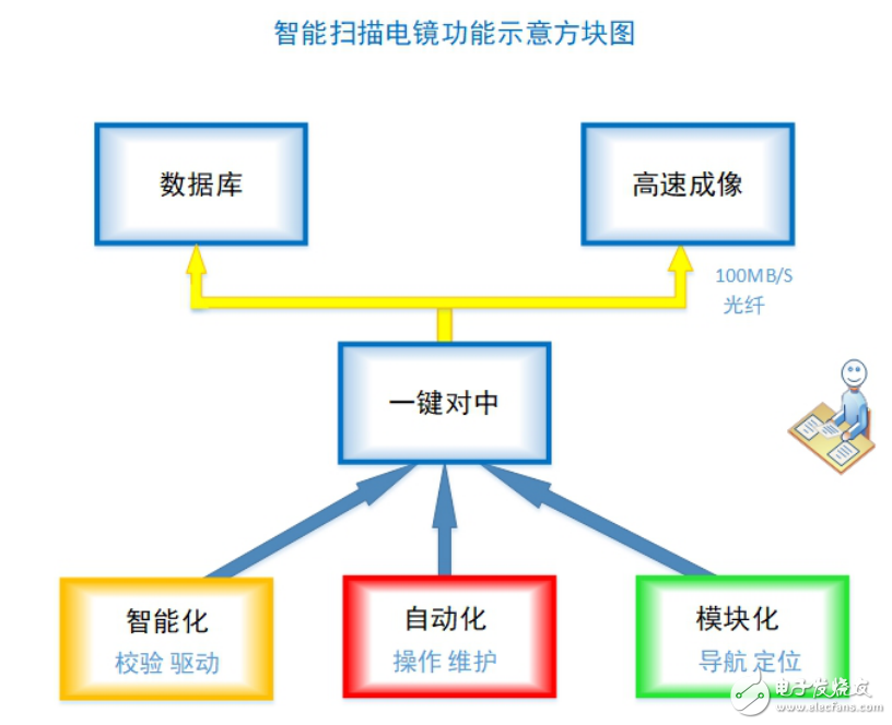
What is a smart electron microscope? The original intention of developing a smart electron microscope is to break through the traditional concept, eliminate misunderstandings, provide a simple environment, and achieve unmanned operation. The schematic diagram of the intelligent electron microscope function module is shown in Figure 1. Specifically, you can use the "five automatic" function to summarize, that is, automatic inspection, automatic feeding, automatic centering, auto focus, automatic operation.

Figure 1 Schematic diagram of the intelligent electron microscope function module
one. Automatic inspectionThe electronic optical system of industrial scanning electron microscope is mainly composed of an electron gun, an electromagnetic lens and a scanning coil. The electron beam is emitted by an electron source, and the thermal field emission electron gun ensures that sufficient initial electrons are emitted onto the sample. The acceleration voltage is set in the range of 0.1 - 12kV. The higher the acceleration voltage, the faster the electron speed, the longer the focal length and the greater the depth of field.
The magnetic field force of the electromagnetic lens does not change the speed of the electron movement, but only changes the direction of electron motion. The high-speed electrostatic deflector is placed in the objective lens, which not only has a compact structure, but also ensures that the aberration of the edge is small when viewed in a large field to achieve high image resolution. The deflector controls the beam to focus on the sample for a quick scan. The detector collects signals such as secondary electrons and backscattered electrons. After the data is processed by amplification, electronic image information can be obtained.
The electro-microscope system is powered on. After the inspection, it will enter the working state. The process of repeated adjustments from top to bottom. Before the electron microscope observation, the optical navigation module will automatically locate, find the ground and navigate, eliminating the need to spend a lot of manual looking for the observation area in front of the mirror, and liberating the engineers from the complicated debugging. The system has a built-in regular full-scale self-test to adjust the electro-optical system to the optimum state of continuous operation.
Automatic inspection is inseparable from the stability system of the support system. The stability of the source plays a primary role. One week of emission current fluctuations, the emission current fluctuation is "1%, which guarantees basic performance.
two. Automatic feedingThe preparation technology is closely matched with the electron microscope, and a large number of samples are continuously observed in the electron microscope, which is one of the keys to improve work efficiency. Taking a biological sample as an example, the biological sample block to be observed is subjected to an epoxy-embedded sample to be hardened and cut, and then cut into a sheet of several tens of nanometers on a microtome. The cut sheets fall into the collecting water tank and are captured by the conveyor belt, and are sequentially collected in a strip arrangement on the round silicon wafer. On the round silicon wafer, dozens or even hundreds of samples can be collected flat, and then the sample is carried into the electron microscope sample stage, and a large number of samples are observed at one time.
The square sample stage with a length of more than 10 cm is freely displaceable along the X-axis and Y-axis precision guide rails. The sample has a repeatability of 200 nm. This avoids the need to reposition each time.
To achieve full automation of electron microscope imaging, it is also necessary to keep up with the preparation technology. The preparation is seamlessly connected with the electron microscope, and the sample is directly sliced ​​and collected, and if necessary, it can be automatically transferred into the electron microscope by means of a strip or a reel.
Round Male Pin Header Connectors
Round Male Pin Header Connectors,Straight Pin Male Pin Header Connectors,Straight Male Pin Header Connectors,Pitch Pin Connector
Dongguan ZhiChuangXing Electronics Co., LTD , https://www.zcxelectronics.com
